
Magnetic Resonance Imaging (MRI) of the prostate is a non-invasive test which can be used to detect, localise and stage prostate cancer. Prostate Cancer is the leading cause of cancer-related deaths in males, killing more than 3,300 men in Australia each year and surpassing the number of deaths caused by breast cancer in women.
Until recently, prostate cancer was difficult to image and detect. MRI has emerged as the imaging modality of choice for evaluating the prostate both before and after cancer has been detected.
Incidence of Prostate Cancer
The risk of prostate cancer based on age is approximately:
Age 40-49 – 1 in 1000
Age 50-59 – 12 in 1000
Age 60-69 – 45 in 1000
Age > 70 – 80 in 1000
although the risk of prostate cancer may increase substantially if there is a family history of prostate cancer.
PSA Tests
A blood test called PSA (prostate-specific antigen) can be performed which if elevated, may indicate the presence of cancer although it may also be elevated by other causes such as benign enlargement of the prostate (called benign prostatic hypertrophy or BPH) or inflammation of the prostate. If the PSA is elevated, a patient may be referred to a specialist urologist or for further evaluation with ultrasound (US) or MRI.
MRI technology and interpretation make a difference
Newer MRI imaging techniques of organs such as the prostate are clearer and more detailed than other imaging methods and enable early cancer detection as well as accurate evaluation of tumour extent (called staging). However, not all MRI machines are the same and the complexity of prostate MRI means that many cancers may be missed by older or less advanced MRI machines. In addition, the skill of the radiographer, who is the trained technician operating the MRI machine and the radiologist, who interprets the images, are paramount in producing the highest quality imaging and the most accurate reporting.
Envision is proud to use the top-of-the-line Siemens 3T Skyra MRI from Germany for our prostate MRIs, the only practice in Perth to do so. This machine, in conjunction with our highly skilled radiographers, produces the clearest and most detailed images of the prostate available which is one reason why more urologists in Perth send their patients to us.
At Envision, prostate scans are interpreted by either Dr Tonya Halliday or Dr Ronny Low who each have more experience in reading prostate MRIs than anyone else in Perth.
MRI-Guided Prostate Biopsy
Following a prostate MRI, specialists may deem a biopsy of the prostate necessary, even if the MRI fails to identify a cancer. This can be done by a urologist using a general anaesthetic and an ultrasound machine to guide needles into the prostate either through the skin (called transperineal biopsy) or through the rectum (called transrectal biopsy). Alternatively, a biopsy of the prostate can be done in an MRI machine using the MRI images to guide the needle into the most suspicious regions (called MRI-guided prostate biopsy or MRGB). Some of the advantages of MRGB include greater accuracy (especially with small lesions) and less trauma to the prostate with many fewer needles inserted into the gland. In addition, MRGB is performed using conscious sedation avoiding the need for a general anaesthetic. A drawback of this technique is that the whole gland can not be sampled.
Dr Tonya Halliday and Dr Ronny Low both perform MRGB at Envision. The biopsies are viewed by pathologist who determines whether cancer is present and if so, how much there is and how aggressive it looks.
Why is MRI a revolution in Prostate Cancer detection and treatment?
For men with intermediate or high probability of spread outside the prostate, MRI improves the accuracy of disease staging. MRI can also assist surgeons plan surgery by showing the extent of the disease enabling a surgeon to preserve delicate arteries and nerves which are essential for maintaining sexual function and bladder control. Whether treatment is necessary and if so, which treatment is best is dependent on many factors including the extent of the tumour.
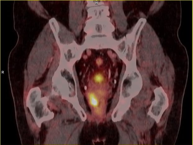
MRI can determine if the cancer is completely inside the prostate gland or has spread to lymph nodes or nearby organs such as the seminal vesicles, bladder, rectum or bones. Additional scans such as a computed tomography (CT), PSMA PET/CT or a bone scan may also be performed to look for cancer outside the prostate.

