What is a Paediatric Hip Ultrasound?

A Paediatric Hip Ultrasound is a procedure that uses ultrasound imaging primarily to evaluate the soft tissues, muscles, tendons, ligaments, joints and bone surrounding the hip in children. It is primarily used in infants to check for developmental dysplasia of the hip (DDH), a condition in which the ball and socket of the hip joint do not form normally.
A Paediatric Hip Ultrasound is used when DDH is suspected or detected, or when there are risk factors such as being born breech or having a family history of DDH.
A series of Paediatric Hip Ultrasounds might be carried out over a period of a few months. This can be to show normal growth of the hip or to show if the abnormality is still present. If your child is having treatment for DDH, further hip ultrasounds might be carried out to detect whether the hip joint is in a good position (for example in a harness) and to show improvement.
Paediatric hip ultrasound studies are also used to look for evidence of fluid in the hip joint. This can indicate joint inflammation, which might be associated with an infection.
Paediatric Hip Ultrasound
What happens during a Paediatric Hip Ultrasound?
A. Before your scan
What to bring
- Your request form
- Any relevant previous imaging
- Your Medicare card and any concession cards
Preparation – the day of your procedure
There is no specific preparation. You will be asked to fill out a questionnaire regarding your child’s health status, medication, and any known allergies. Your child will need to have their nappy and clothing removed to allow the ultrasound study to be carried out.
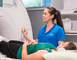
B. During your Paediatric Hip Ultrasound
Procedure
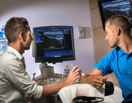
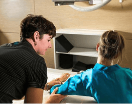
Your child will be placed on their side or back on an ultrasound bed, and their knees will usually be bent during the scan. Gel will be applied to the area being imaged to help create a good contact between you and the ultrasound probe. The probe will be placed directly onto the gel and your skin for the duration of the examination.
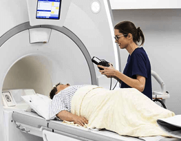
Your ultrasound will be performed by a Radiologist (medical specialist) or a sonographer (a specially trained technologist). Because the examiner is interpreting moving images on a screen a high degree of concentration is required.
Ultrasound examinations are not painful and generally not invasive but may be uncomfortable particularly if you need to move a body part that causes you discomfort.
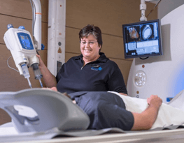
Most ultrasound examinations will be completed within 30 minutes. It is not unusual for the radiologist to come in and speak with you and view the images on the screen. At the end of the procedure the gel is simply wiped from your skin so that it does not mark your clothes.
Risks and side effects
Ultrasounds are a very low risk procedure and complications are rare however you should be informed of the possible risks and side effects.
Risks associated with this procedure include:
- If scanning is performed over an area of tenderness, you may feel pressure or minor pain from the transducer.
Any medical procedure can potentially be associated with unpredictable risks.
Who will perform the Paediatric Hip Ultrasound?
Your ultrasound examination will be carried out by a sonographer (a technologist trained in ultrasound imaging)
Paediatric Hip Ultrasound
What happens after a Paediatric Hip Ultrasound?
How do I get my results?
After your child’s appointment, the information from your scan is interpreted by Envision’s Radiologist before delivery of a report to your child’s doctor or Paediatrician.
Post-procedure
Your child should be able to go about your daily activities after your appointment.
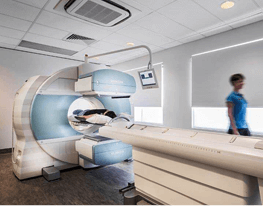
Medical Imaging Practice Perth
Types of Imaging
At Envision, we offer the most sought-after types of imaging for diagnostics and treatments. Our Wembley headquarters is the largest single-site radiology practice in Perth